Fig. 5.

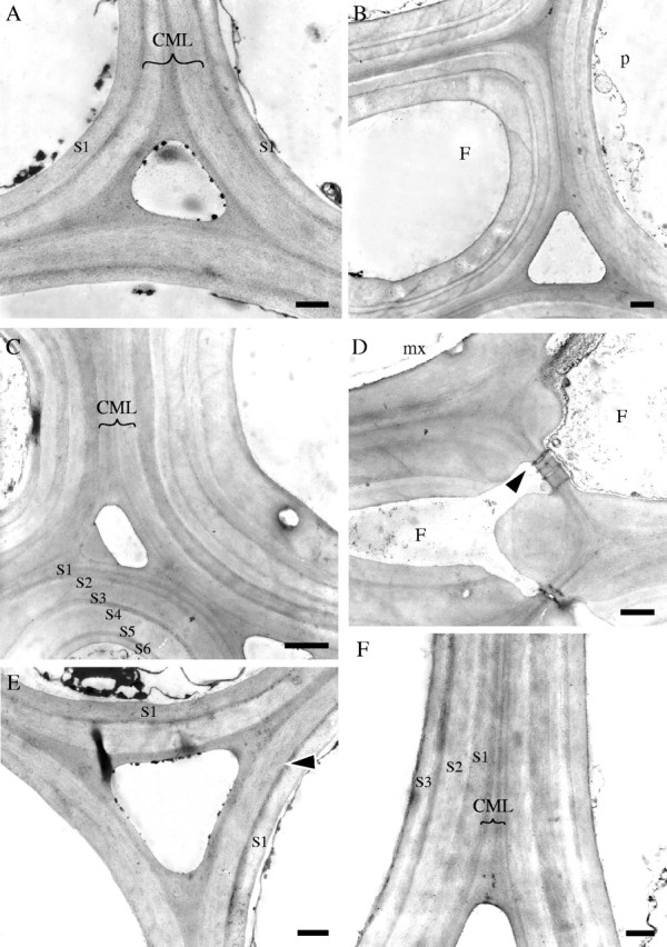
Stage 4 of development (internode 1 of immature and mature shoots). TEM micrographs (all TS). (A) Cell walls of three fibres from the centre of protoxylem sheath at internode 1 of the immature shoot showing compound middle lamella (CML), a thin dark transition layer, and one layer of secondary wall (S1). (B) Fibre (F) from the middle of the protoxylem sheath located next to a parenchyma cell (p) at the periphery of the sheath (internode 1, mature shoot). At least three secondary wall layers can be recognized. Note variation in the thickness of individual layers. (C) Cell walls of fibres three cells away from a metaxylem vessel (mature shoot). At least six secondary wall layers can be distinguished (S1–S6). (D) Cell walls of flattened fibres (F) adjacent to a metaxylem vessel (mx) showing wall layering with less clear boundaries. Plasmodesmata connect pits between two cells (arrow) (mature shoot). (E, F) Parenchyma cell walls. In the immature shoot (E) only a thin dark boundary layer (arrow) and one secondary layer (S1) have been deposited, whereas in the mature shoot (F) further layers (S1–S3) have been deposited. Scale bars: (A, D–F) 0·5 μm, (B, C) 1·0 μm.
