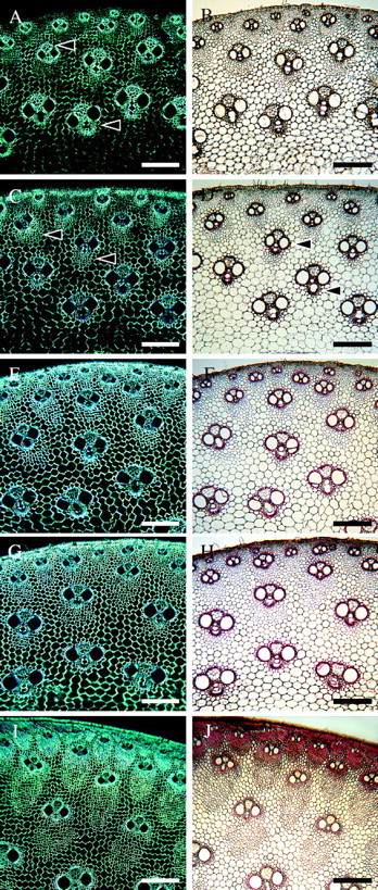
Fig. 7.

Stages 3 and 4 of tissue development as observed with polarized light (A, C, E, G, I) and with phloroglucinol–HCl staining (B, D, F, H, J). (A, B) Internode 5. Birefringence is pronounced mainly in fibres adjacent to the vascular elements (arrows), indicating presence of secondary cell walls. Cell walls remain unlignified (B). (C, D) Internode 4. Increase in birefringence in fibres not adjacent to vascular elements (arrows). Lignification starts in fibres surrounding the vascular tissues (arrows in D). (E, F) Internode 3. All cell walls become birefringent, including parenchyma. Lignification is still mainly confined to vascular tissues and cells close to their vicinity; however, staining is more intense. (G, H) Internode 2, immature shoot. All cell walls are highly birefringent. Phloroglucinol staining is slightly more pronounced than in the previous internode (H). (I, J) Internode 2, mature shoot. Further secondary wall deposition is evident due to the high level of birefringence. Cell walls continue to lignify in an inward direction. Scale bars: all 200 μm.
