Fig. 2.

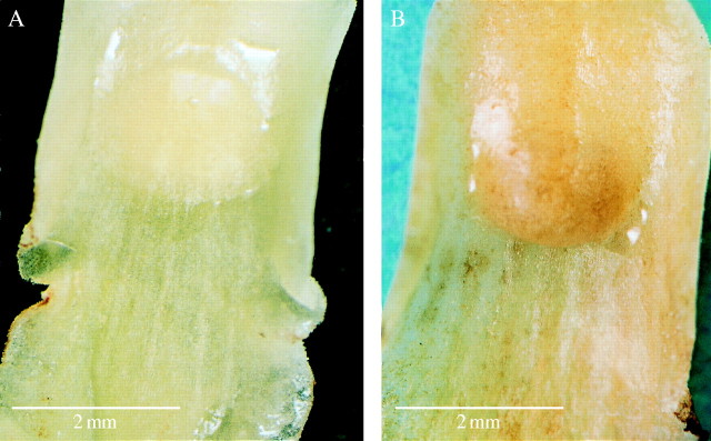
(A) Labellar surface during early anthesis showing simple callus and associated drops of nectar. Scale bar = 2 mm. (B). Labellar surface at day 4 with nectar accumulating at distal end of callus. Scale bar = 2 mm.

(A) Labellar surface during early anthesis showing simple callus and associated drops of nectar. Scale bar = 2 mm. (B). Labellar surface at day 4 with nectar accumulating at distal end of callus. Scale bar = 2 mm.