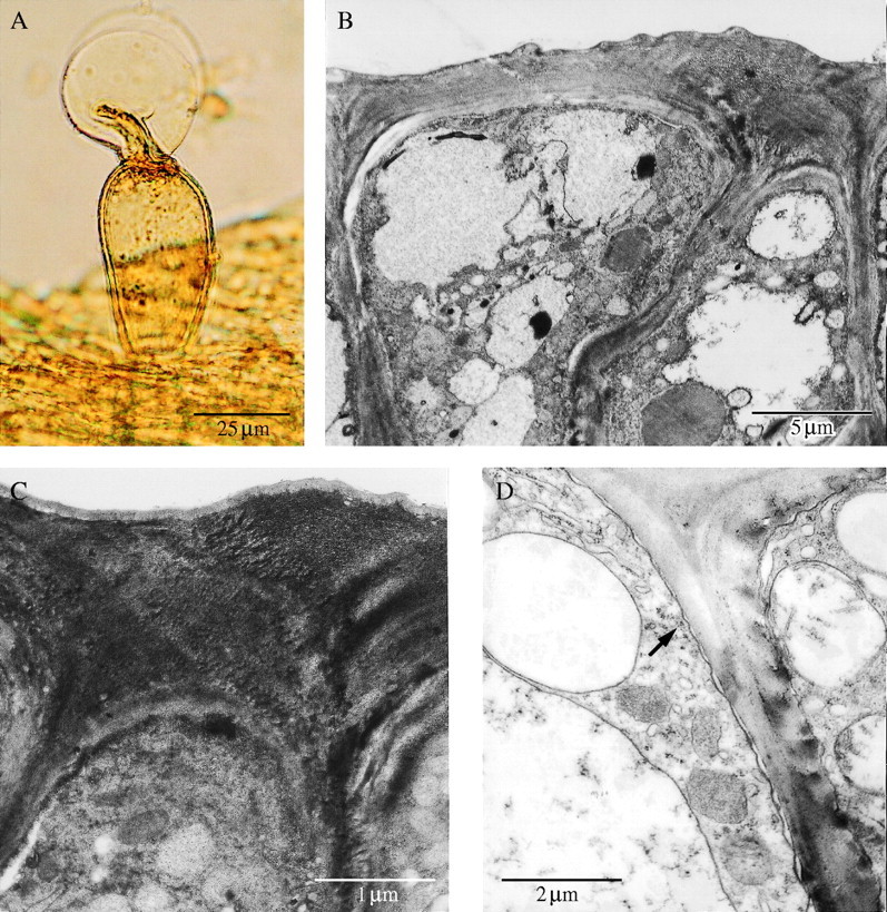
Fig. 5.

(A) Stalked, glandular trichome on surface of labellar callus with exudate that possibly contains terpenes. Scale bar = 25 µm. (B, C) TEM of cells of secretory epidermis showing thick, outer, tangential wall and uninterrupted cuticle. Scale bars = 5 µm and 1 µm, respectively. (D) Secretory callus tissue during bud stage showing mitochondria, rough ER, components of the vacuome, tonoplast and coated vesicles (arrow). Scale bar = 2 µm.
