Fig. 6.

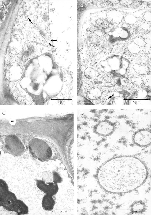
(A, B) Abaxial, subepidermal, labellar cells with mitochondria, rough ER, amyloplasts with starch grains, dictyosomes, components of the vacuole and coated vesicles (arrows). Scale bars = 2 µm and 5 µm, respectively. (C) Secretory, epidermal cell 4 d into anthesis showing outer tangential wall (w), parietal cytoplasm and much of the cell volume occupied by a vacuole. Note the plastids and intravacuolar, osmiophilic bodies and compare them with those found in cells at the early stages of flowering (Fig. 5B). In both cases, the plastids have a homogeneous matrix but few plastoglobuli and lamellae, whereas 4 d into anthesis, the osmiophilic bodies are larger and occur more frequently. Scale bar = 2 µm. (D) Secretory cells 4 d into anthesis showing intravacuolar, annular profiles. Scale bar = 2 µm.
