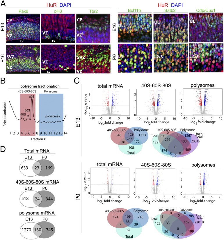
Fig. 1.
HuR regulates mRNA translation in mitotic neural stem cells and differentiating projection neurons of the developing neocortex. (A) Representative coronal confocal images of immunostained developing neocortices. HuR immunohistochemistry (red) shows that HuR is expressed in RG neural progenitors colabeled with Pax6 (green) and pH3 (green) at E13 and E16 in the ventricular zone (VZ). HuR is also expressed in postmitotic differentiated Bcl11b-positive lower-layer neurons (green) and in upper-layer neurons labeled with Satb2 and Cdp/Cux1 (green) at E16 and P0. CP, cortical plate; DL, deep layers; L2/3, layer 2/3; L5, layer 5; LV, lateral ventricle; SVZ, subventricular zone; UL, upper layers. (B) Schematic of sucrose density gradient fractionation and isolation of 40S–60S–80S and polysome cytoplasmic components for analysis. (C) E13 and P0 WT and Emx1–HuR-cKO neocortices were fractionated into 40S–60S–80S and polysomes, then subjected to RNAseq coupled with bioinformatics analysis. Volcano plots show gene-expression levels relative to WT; blue dots represent higher expression in HuR-cKO; red dots represent lower expression in HuR-cKO; and gray dots represent unchanged levels at a false-discovery rate ≤5%. Venn diagrams show the number of genes that change with HuR-cKO in 40S–60S–80S and polysomal fractions, with respect to total mRNA expression levels (whether changed or unchanged), analyzed by RNAseq at E13 and P0. (D) Venn diagrams show total, 40S–60S–80S, and polysome-associated mRNAs that change in abundance in response to HuR deletion. The mRNAs are unique to E13, unique to P0, or present at both developmental stages.

