Fig. 3.
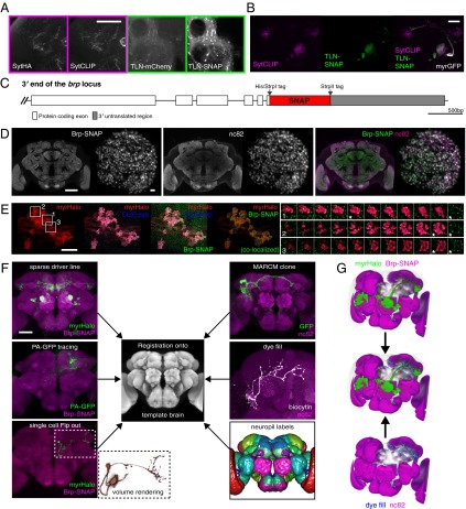
Key applications for chemical labeling in Drosophila neurobiology. (A) Comparison between epitope-tagged synaptic markers (HA-tag, mCherry) and synaptically targeted chemical tags (SNAP-tag, CLIP-tag). Confocal slices from the brain of a fruGal4> SytHA, SytCLIP (presynaptic markers) animal (Left) and a fruGal4> TLN-mCherry (DenMark), TLN-SNAP (somatodendritic markers) animal (Right) are shown. (Scale bar: 50 µm.) (B) Simultaneous labeling of presynaptic sites and the somatodendritic compartment of DA1 projections neurons using Mz19Gal4. (Scale bar: 50 µm.) (C) Map of the brp-SNAP knockin. (D) nc82 and Brp-SNAP signals colocalize in a brp-SNAP/+ brain simultaneously labeled with BG-549 and nc82 immunostaining. Single coronal slices through the middle of the brain (Left) and through a deconvolved image stack of the DA1 glomerulus (Right) are shown. Note the more even Brp-SNAP staining in the center of the brain. (Scale bars: 50 µm, Left; 2 µm, Right.) (E) Deconvolved, confocal z-stack images from the MB calyx region of a brp-SNAP, Mz19-Gal4 > CLIP-syb, myrHalo brain triple-labeled with fluorescent BG-488, BC-547, and Halo-SiR substrates. Colocalization analysis of SNAP-tag– and Halo-tag–labeled regions reveals presynaptic sites (Brp puncta) within PN terminals (see also Movie S1). (Scale bar: 10 µm.) Montages show three individual, triple-labeled projection neuron terminals (Right, 1–3) derived from the indicated boxed regions. One selected optical section (marked with an asterisk) is also shown with Brp-SNAP only (green), revealing that puncta are localized to the surface of each terminal. (F) Chemically labeled neurons can be registered successfully onto a template using the Brp-SNAP neuropil counterstaining, thus allowing direct comparison with imaged neurons from other sources, such as those derived from stochastic labeling (Flp-out), PA-GFP tracing, whole-cell recordings, or sparse driver lines, (e.g., Mz19-Gal4). (Scale bar: 50 µm.) (G) Overlay of DA1 projection neurons (green) from a chemically labeled brp-SNAP, Mz19-Gal4 > myrHalo brain (Left) overlaid with a dye-filled third-order olfactory neuron (cyan) from an nc82-stained brain (Right) after registration. Note the overlap between axon terminals and dendritic arbor (white arrowheads).

