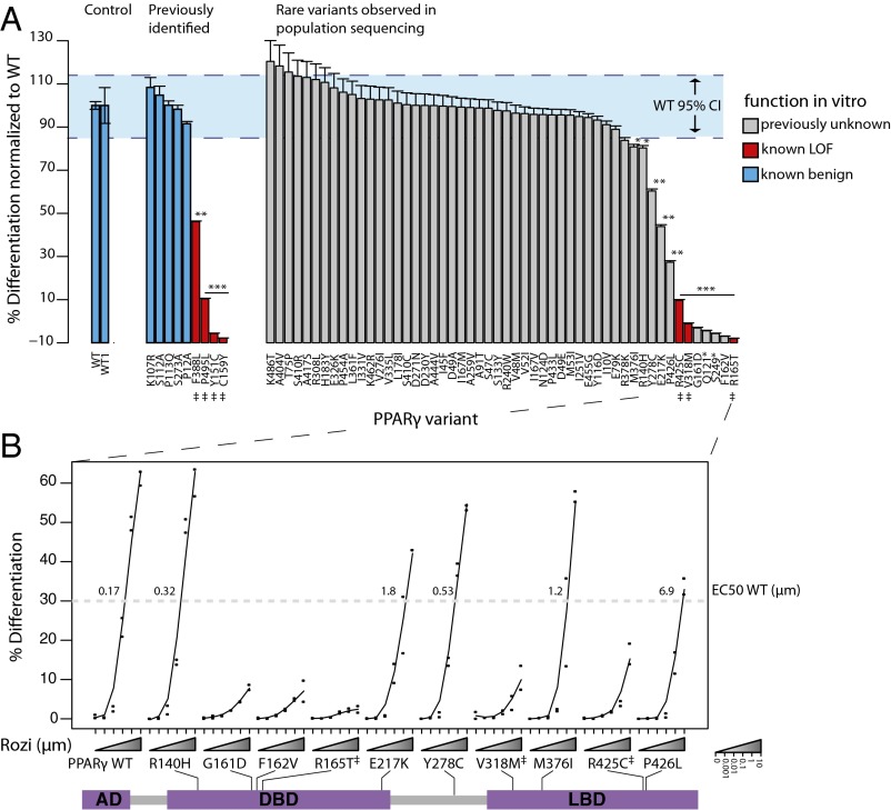
Fig. 2.
Experimental characterization of rare PPARγ variants identified from population sequencing. (A) Each PPARγ variant was generated and tested for its ability to rescue adipocyte differentiation in vitro. From left to right PPARγ variants are sorted by in vitro function in three groups: (i) WT from independent experiments, (ii) previously identified synthetic and human mutations, and (iii) variants identified in population based exon resequencing. Blue dashed lines denote the 95% confidence interval of WT function. (B) Roziglitazone (rozi) dose–response of PPARγ variants identified as LOF. The amino acid position along the PPARγ protein is shown. EC50 WT denotes the rozi dose required to achieve 50% of maximal WT response. AD, activation domain; DBD, DNA-binding domain; LBD, ligand-binding domain. Error bars indicate ±1 SEM. Significant differences compared with WT are noted: *P < 0.05; **P < 0.005; ***P < 0.0001. ‡Variants identified in families with partial lipodystrophy.

