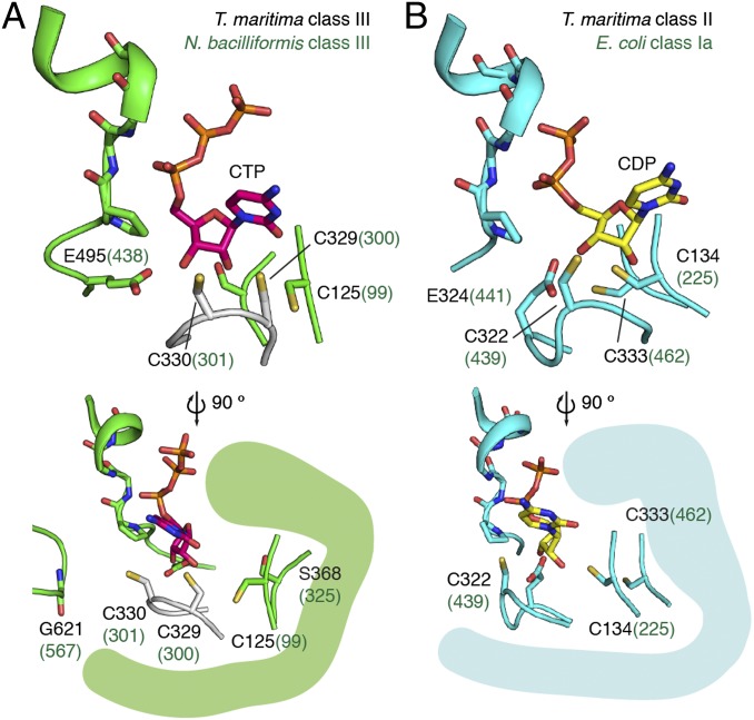
Fig. 6.
Model for disulfide formation in class III RNR. (A) The crystal structure of TmNrdD is shown in green (the inserted loop, residues 350–365, is not shown for clarity). The Cys loop based on bacteriophage T4 NrdD (PDB ID code 1H79) (45) has been modeled in white. CTP has been modeled based on the T maritima class II RNR structure (magenta). The side view shows the position of the G• loop. (B) The structure of CDP (yellow) bound T. maritima class II RNR (PDB ID code 1XJN) (46).

