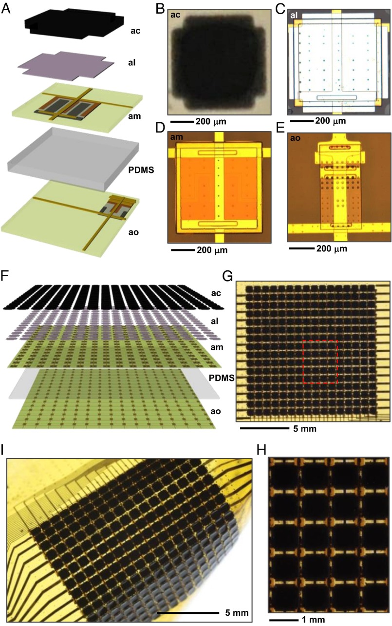
Fig. 1.
Schematic illustrations and images of adaptive camouflage systems that incorporate essential design features found in the skins of cephalopods. (A) Exploded view illustration of a single unit cell that highlights the different components and the multilayer architecture. The first and second layers from the top correspond to the leucodye composite (artificial chromatophore; ac) and the Ag white reflective background (artificial leucodye; al). The third layer supports an ultrathin silicon diode for actuation, with a role analogous to that of the muscle fibers that modulate the cephalopod’s chromatophore (artificial muscle; am). The bottom layer, separated from the third layer by PDMS, provides distributed, multiplexed photodetection, similar to a postulated function of opsin proteins found throughout the cephalopod skin (artificial opsin; ao). (B) Optical image of a thermochromic equivalent to a chromatophore. (C) Optical micrograph of the Ag layer and the silicon diode. (D) Optical image of the diode. (E) Optical image of a photodiode and an associated blocking diode for multiplexing. (F) Exploded view of illustration of a 16 × 16 array of interconnected unit cells in a full, adaptive camouflage skin. (G) Top view image of such a device. (H) Image of the region highlighted by the red box in G. (I) Image of a device in a bent configuration.

