Fig. 4.
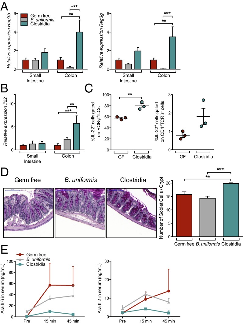
Clostridia colonization induces IL-22. (A) Reg3b and Reg3g expression from whole-tissue extracts isolated 4 d after colonization from the small intestine or colon of GF (red), B. uniformis-colonized (gray), or Clostridia-colonized (green) mice. Quantitative RT-PCR data are plotted relative to GF and normalized to Hprt (n = 8–9 mice per group from two independent experiments). (B) Il22 expression in LPL from mice in A. (C) IL-22 production by RORγt+ ILCs and T cells 6 d after colonization, determined by flow cytometric analysis of permeabilized cells (SI Methods; n = 3 mice per group representative of three independent experiments). (D) Representative images and quantification of goblet cells in distal colon of GF, B. uniformis-colonized, and Clostridia-colonized mice 6 d after colonization. n = 3–5 mice per group. (Scale bar, 100 µm.) (E) Serum Ara h 6 and Ara h 2 levels after PN gavage in GF, B. uniformis-colonized, or Clostridia-colonized mice 6 d after colonization (n = 5–12 mice per group from two independent experiments). *P < 0.05, **P < 0.01, ***P < 0.001 by two-way ANOVA with Bonferroni posttest (A and B) or one-way ANOVA with Tukey posttest (C).

