Fig. 4.
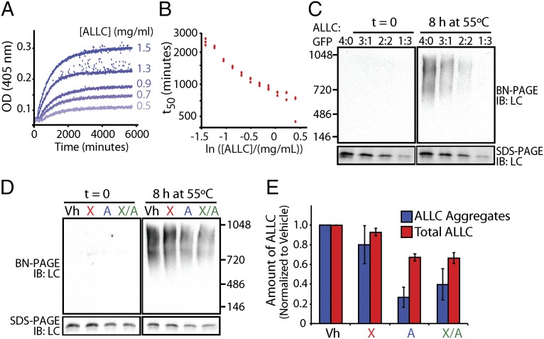
Stress-independent activation of XBP1s and especially ATF6 reduces aggregation of cell-secreted ALLC. (A) Timecourses of recombinant ALLC aggregation at the indicated concentrations. Samples were incubated at 44 °C for the indicated time, and aggregation was measured by turbidity at 405 nm. (B) Plot of the t50 of recombinant ALLC aggregation at the indicated concentration from data as shown in A; n = 3 replicates are shown. (C) Immunoblots for BN-PAGE and SDS-PAGE of media conditioned on HEK293DAX cells expressing FTALLC for 24 h. The media was diluted with media conditioned on GFP-expressing cells, as indicated, and incubated at 55 °C for 8 h. (D) Representative immunoblots for BN-PAGE and SDS-PAGE of media conditioned on HEK293DAX cells following a 16-h preactivation of XBP1s (X), ATF6 (A), or XBP1s and ATF6 (X/A), as in Fig. 3B. FTALLC aggregation was induced by incubating the media for 8 h at 55 °C. (E) Graph depicting the quantification of soluble aggregates and total ALLC from BN-PAGE and SDS-PAGE immunoblots, as shown in D, normalized to vehicle. Error bars represent SEM from biological replicates (n = 3).

