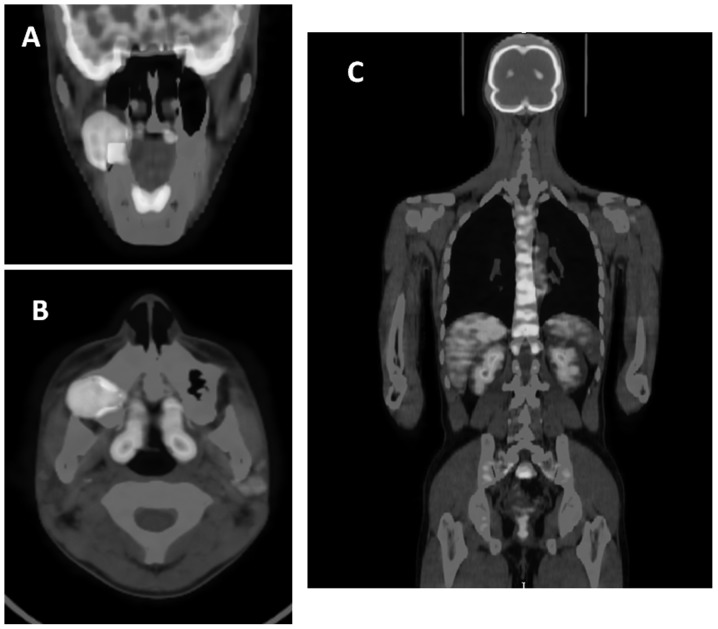
Figure 4.
FDG-PET images. (A) axial and (B) coronal FDG-PET/CT images revealing a slight FDG uptake in the primary tumor of the right maxilla and bilateral superior internal jugular nodes. (C) No abnormal uptake, which would indicate distant metastasis, was observed on FDG-PET images. FDG-PET, 18F-fluorodeoxyglucose-positron emission tomography; CT, computed tomography.

