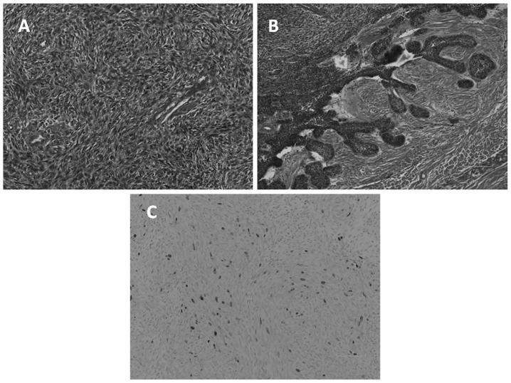
Figure 5.
Microscopic examination. (A) The majority of the mass consisted of spindle tumor cells exhibiting a storiform, pseudosarcomatous pattern. The epithelial component demonstrated cytological malignancy, characterized by nuclear pleomorphism, an increased nucleus to cytoplasm ratio, hyperchromatic nuclei and a high mitotic rate. (B) In the other area, the tumor cell nest exhibited peripheral palisading of columnar cells, with a vacuolated cytoplasm and reverse-polarized nuclei. These findings resemble those for ameloblastoma. (C) The Ki-67 proliferation index was 5%, indicating that this tumor was of low malignancy.

