
Figure 4.
Case two: (A) Gastroduodenoscopy demonstrating a large ulcer of the antrum (arrow) and (B) hematoxylin and eosin staining of the lesion revealing a primary malignant gastric lymphoma confined to the mucosa (circled; magnification, ×400).
Official websites use .gov
A
.gov website belongs to an official
government organization in the United States.
Secure .gov websites use HTTPS
A lock (
) or https:// means you've safely
connected to the .gov website. Share sensitive
information only on official, secure websites.
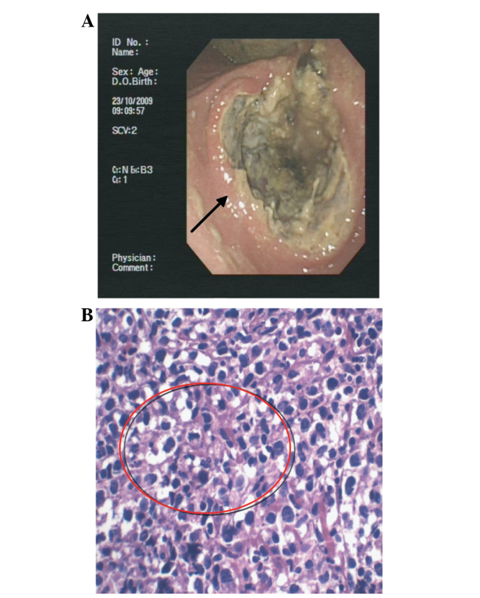
Case two: (A) Gastroduodenoscopy demonstrating a large ulcer of the antrum (arrow) and (B) hematoxylin and eosin staining of the lesion revealing a primary malignant gastric lymphoma confined to the mucosa (circled; magnification, ×400).