Figure 5.

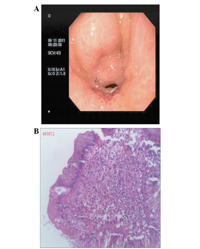
(A) Gastroduodenoscopy demonstrating an ulcer on the pyloric canal and (B) hematoxylin and eosin staining of the lesion revealing it to be an ulcer accompanied by inflammation (magnification, ×100).

(A) Gastroduodenoscopy demonstrating an ulcer on the pyloric canal and (B) hematoxylin and eosin staining of the lesion revealing it to be an ulcer accompanied by inflammation (magnification, ×100).