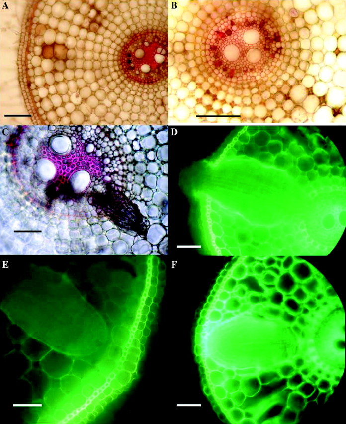
Fig. 6.

Rice: effects of 2 d in 0·174 mm sulfide on root anatomy, 2 weeks after lifting of treatment. Fresh hand-cut transverse sections of adventitious roots; (A), (B) and (C) stained with phloroglucinol and concentrated hydrochloric acid to show lignification (red); (D), (E) and (F) viewed with blue light to show yellow autofluorescence of lipid material in cell walls. (A) Sulfide treatment, 15 mm from the apex showing heavy lignification of stele, vascular blockages, brown occlusions in cortical intercellular spaces and slight lignification of exodermis. None of these blockages or lignifications was seen in the controls. Bar = 50 µm. (B) High power of stele similar to (A), showing lignified blockages of protoxylem (red) and blocked phloem (brown). Bar = 50 µm. (C) Sulfide treatment 30 mm from the apex, showing a lateral root which had died prior to emergence. Bar = 50 µm. (No such death of laterals was seen in the controls.) (D) Sulfide treatment, 40 mm from the apex showing emergent lateral root swollen and fluorescing within the adventitious root cortex; control laterals did not become swollen or fluoresce. Bar = 50 µm. (E) Control, 30 mm from the apex, with lateral root prior to emergence; note that opposite the lateral is a ‘window’ in the hypodermis and exodermis with virtually no thickening or autofluorescence (cf. F). Bar = 50 µm. (F) Sulfide treatment, 30 mm from the apex, with lateral root, showing thickened exodermis and strong autofluorescence of adventitious root exo/hypodermis opposite the lateral, and of the edge of the lateral (cf. E). Bar = 50 µm.
