Fig. 7.

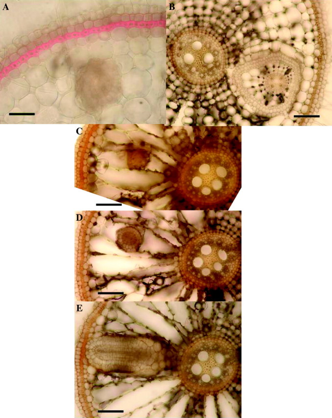
Rice: effects of 2 d in 0·174 mm sulfide on lateral root growth, 2 weeks after lifting of treatment. Fresh hand-cut transverse sections of adventitious roots (length = 100–120 mm); (A) stained with phloroglucinol and concentrated hydrochloric acid to show lignification (red); (B–E) unstained. (A) Approx. 45 mm from the apex, showing strongly thickened and lignified exodermis and lateral root growing through the cortex. (Control roots had only slightly lignified exodermis.) Bar = 50 µm. (B) Approx 40 mm from the apex, showing a single lateral growing through adventitious root cortex; note that the lateral has enlarged cortical cells and gas spaces and hypodermal differentiation. Bar = 50 µm. (C), (D) and (E) are serial sections of one root from the base towards the apex, taken 90–95 mm from the apex; (C) is near the tip of the lateral, while (E) is near the basal part which connects with the stele of the adventitious root. The laterals were therefore growing upwards through the adventitious root cortex. Bars = 50 µm. (No control roots were found with laterals growing within the parent root cortex; here laterals emerged normally.)
