
Fig. 8.
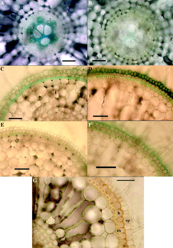
Rice: evidence for sulfide-induced barrier to Fe2+ absorption in adventitious roots. (A–F) Effects of 2 d of 0·174 mm sulfide on Fe2+ absorption, 2 weeks after lifting of treatment. Excised roots (length = 100–120 mm); iron indicated as Prussian blue precipitate of Fe4(Fe(CN)6)3. (A) Control, root base: appreciable iron present in the stele. Bar = 50 µm. (B) Sulfide treatment, root base: comparatively little iron present in the stele (cf. A). Bar = 50 µm. (C) Control, 25 mm from the apex: hypodermal layers do not appear to be great barriers to Fe2+ (cf. D). Bar = 50 µm. (D) Sulfide treatment, 25 mm from the apex: Fe2+ accumulated in the epidermis and hypodermal layer appears to be the barrier to Fe2+ (cf. C and G). Bar = 50 µm. (E) Control, 5 mm from the apex: hypodermal layers apparently formed little barrier to Fe2+ (cf. F). Bar = 50 µm. (F) Sulfide treatment, 5 mm from the apex: Fe2+ accumulated in epidermis; the hypodermal layer appears to be the barrier to Fe2+ (cf. D, E and G). Bar = 50 µm. (G) Transverse section of the base of a root stained in iodine from sulfide treatment, but which had not been in an Fe2+ absorption experiment. Note the thickening of outer tangential and radial walls of the hypodermal layer which appear to form a barrier (cf. D and F). Bar = 50 µm. Ep = epidermis; h = hypodermis; ex = exodermis.