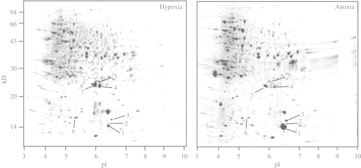
Fig. 1.
Patterns of proteins in 7–11 mm tips of coleoptile dissected from intact rice seedlings before and after 72 h in anoxia. Data are Coomassie blue G-250-stained coleoptile proteins (600 µg per gel) separated by two-dimensional IEF/SDS–PAGE. Seedlings were germinated and grown for 48 h in aerated solution (0·25 mm O2), then pre-treated with hypoxia (0·028 mm O2) for 16 h prior to 72 h anoxia. Coleoptile tips contained no leaf tissues. A gel showing the pattern of proteins for aerated coleoptiles can be seen in supplementary Fig. 1. It is similar to that shown above for coleoptiles treated hypoxically for 16 h.

