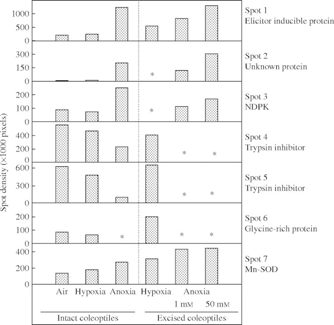
Fig. 2.
Changes of spot density in gels for protein separation of samples from rice coleoptile tips, after 72 h anoxia. Protein was extracted from coleoptile tips (top 7–11 mm), either dissected after intact seedlings had been exposed to anoxia, or from tips excised and healed before anoxia was imposed, and then supplied with either 1 or 50 mm exogenous glucose. Data are derived from supplementary Fig. 1. An asterisk indicates that the spot could not be analysed due to low abundance.

