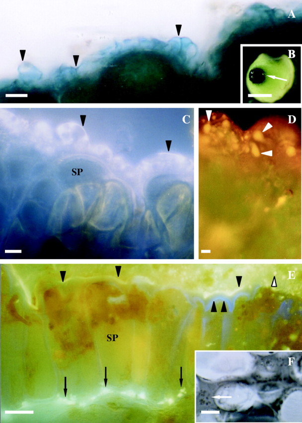
Fig. 2.

Histochemistry of stigmatic region of Sarcandra glabra at receptivity. (A) Arrowheads mark dark blue-positive esterase at cuticle surface of stigmatic epidermal cells. Free-hand section of fresh tissue. (B) Low magnification view of esterase-positive stigma (arrow). (C) 8-ANS-positive fluorescence (arrowheads) indicating protein distribution at the cuticle proper and cuticular layer of the stigmatic epidermal cell. Fresh tissue whole mount. (D) Orange nile-red-positive fluorescence demonstrating presence of neutral lipids (arrowheads) throughout the cuticle proper and cuticular layer of stigmatic epidermal ECM. Fresh tissue whole mount. (E) Auramine-O-positive yellow-green fluorescence (arrowheads) showing presence of culticle. Arrows denote lipids in expanded ECM between stigmatic epidermal cells and adjacent cells where pollen tubes will grow. Double arrowhead marks primary wall. White arrowhead notes auramine-O-positive droplet. Free-hand section of fresh tissue. (F) Arrow marks osmiophilic-positive droplets in expanded ECM between stigmatic epidermal cells and subtending cells where pollen tubes will grow. Cryofix, serial section. Scale bars: B = 7 mm; A, C–F = 10 µm. SP, Stigmatic papilla.
