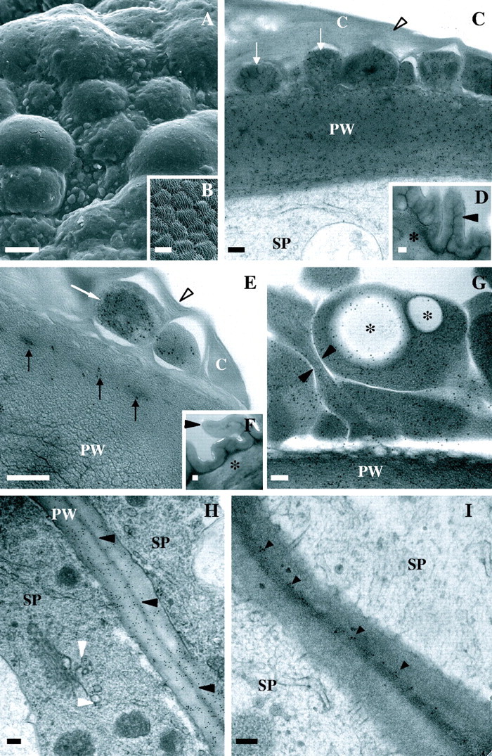
Fig. 3.

Scanning and transmission electron micrographs of stigmatic and abaxial epidermal cells of Sarcandra glabra. (C–I) Immunolocalization of anti-JIM7 or JIM5 (18 nm gold particles). (A) Heterogenous surface topology of stigmatic ECM. (B) Ridged surface topology of abaxial epidermis. (C) Anti-JIM7 in cuticular layer (arrows) and subtending primary wall of stigmatic epidermal cells. Arrowhead marks thinly lamellate cuticle proper. (D) Anti-JIM7 in primary wall of abaxial epidermal cell. Arrowhead and asterisk mark thick cuticle and primary wall, respectively. (E) Ant-JIM in cuticular layer (arrows) and primary wall of stigmatic epidermal cells. Arowhead denotes thinly lamellate cuticle. (F) Anti-JIM5 in primary wall of abaxial epidermal cell. Arrowhead and asterisk mark thick cuticle and primary wall, respectively. (G) Anti-JIM7 in cuticular layer arrows and primary wall of stigmatic epidermal cell. Note large spherical immunonegative spherical regions (asterisks) which are likely to correspond to nile-red- and auramino-O-positive regions. Arrowheads mark channels separating homogalacturonan-rich areas within the cuticular layer. (H) Anti-JIM7 between stigmatic epidermal cells. Black arrowheads mark middle lamella. White arrowheads denote gold particles in Golgi body-associated vesicles. (I) Anti-JIM5 in middle lamella (arrowheads) between stigmatic epidermal cells. Scale bars: A and B = 10 µm; C–I = 200 nm. C, Cuticle; PW, primary wall; SP, stigmatic papilla.
