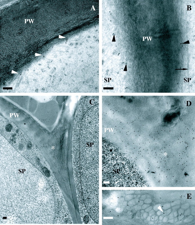
Fig. 4.

Immunolocalization of JIM13 (18 nm gold particles; A and B) and SCA (18 nm gold particles; C–E) in stigmatic epidermal cell ECM. (A) Localization at plasma membrane/primary wall boundary (arrowheads) at surface of stigmatic epidermal cells. (B) Localization at plasma membrane/primary wall boundary (arrowheads) between stigmatic epidermal cells. Arrow marks microtubule. (C and D) Localization in primary wall at notch (asterisk) between stigmatic epidermal cells. (E) Localization in expanded ECM in association with putative auramine-O/nile red-positive droplets (arrowhead). Scale bars = 200 nm; PW, primary wall; SP, stigmatic papilla.
