
Fig. 6.
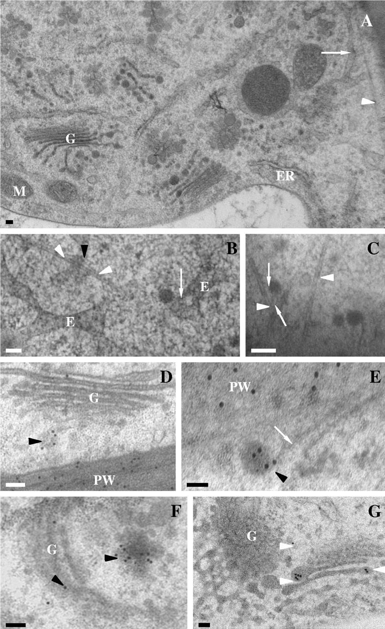
Stigmatic epidermal cell ultrastructure in Sarcandra glabra. (A) Low magnification illustrating extensive endomembrane system. Arrow denotes tubular-vesicular endosome-like array. Arrowhead marks microtubule. (B) Tubular-vesicular endosomal-like array with tethered vesicles (arrow) and microtubule (arrowheads). (C) Vesicles tethered (arrows) to microtubules (arrowheads). (D–F) Immunolocalization of JIM7 (18-nm gold particles) to Golgi body-associated vesicle (black arrowheads). White arrow marks microtubule. (G) Immunolocalization of SCA (18-nm gold particles) to Golgi body-associated vesicles (arrowheads). Scale bars = 100 nm. E, Tubular-vesicular endosome-like array; ER, endoplasmic reticulum; G, Golgi body; PW, primary wall.