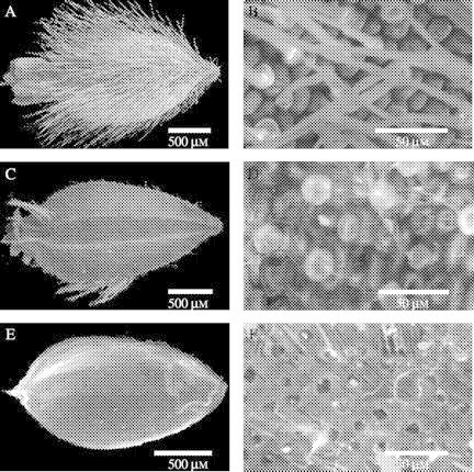
Fig. 6.

ESEM images of the surface of Actinotus leucocephalus seeds following laboratory storage (A,B), burial in the unburnt site for 24 months (C,D) and surface sterilization following burial in the unburnt site for 18 months (E,F). For each pair of images the whole dispersal unit is shown (A,C,E) and then the mid-section of the seeds is captured at higher magnification (images B, D and F, respectively).
