
Fig. 4.
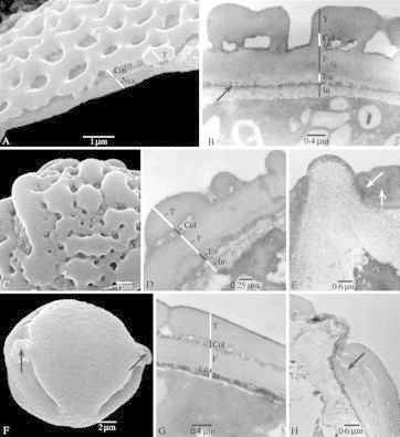
SEM and TEM pictures showing the surfaces and sections of the pollen wall in Marcgravia and Souroubea. (A and C) treated with acetolysis; (B and D–H) critical point dried. (A) Marcgravia macrophylla, SEM, thin pollen wall showing nexine (Nex), columellae (Col) and tectum (T). (B) M. rectiflora (Fuertes 587), TEM, thin pollen wall showing intine (In), endexine (En), foot layer (F), columellae (Col) and tectum (T); the arrow points to granular endexine. (C) M. nervosa (Maas and Dressler 746), SEM, detail of perforate–verrucate sexine ornamentation. (D) M. nervosa (Stevens 23729), TEM, ultrastructure of pollen wall showing intine (In), endexine (En), foot layer (F), columellae (Col) and tectum (T). (E) M. nervosa (Stevens 23729), TEM, protruding intine at the apertural region, endexine lamellated near aperture (arrows). (F) Souroubea guianensis subsp. cylindrica, SEM, equatorial overview showing papillae (arrows). (G) S. sympetala (Smith 1555), TEM, detail of pollen wall showing intine (In), endexine (En), foot layer (F), columellae (Col) and tectum (T). (H) S. guianensis subsp. guianensis (Gillespie 1200), TEM, papillae consisting of protruding intine at the apertural region, endexine lamellated near apertures (arrow).