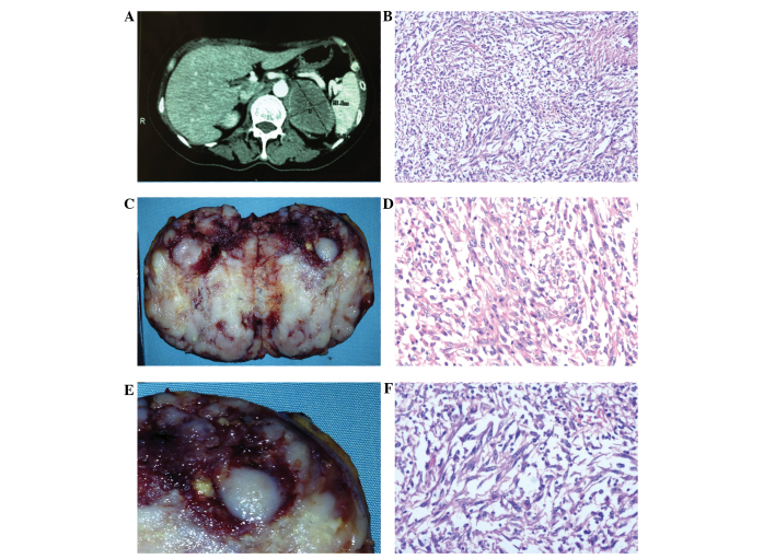
Figure 1.
Gross images and hematoxylin and eosin (H&E)-stained microscopic images of the primary adrenocortical carcinosarcoma. (A) A computed tomography scan demonstrates a 7.6×5.1-cm well-demarcated and peripherally enhanced left adrenal mass impinging on the pancreas and the spleen, without parenchymal invasion into the kidney. (B) Gross image of the cut surface of the adrenocortical carcinosarcoma shows a circumscribed mass, predominantly comprising paler, firm tissue, with partially yellow tissue. (C) A compressed rim of normal adrenal gland is apparent adjacent to the tumor capsule. (D) The tumor comprises of spindle cell and epithelial components (H&E staining; magnification, ×100). (E) Carcinomatous cells exhibit highly atypical nuclei and an abundant eosinophilic cytoplasm (H&E staining; magnification, ×200). (F) Spindle-shaped tumor cells reveal highly pleomorphic nuclei (H&E staining; magnification, ×200).

