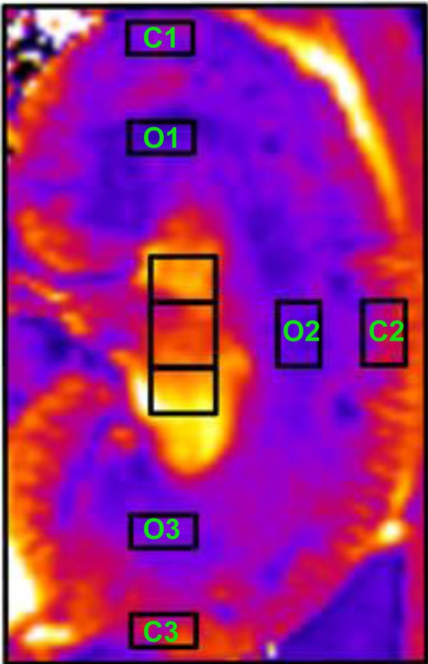
Figure 4.

Selection of regions of interest. C 1–3 identify cortical regions of interest. O 1–3 identify outer medullary regions of interest.
Notes: Adapted from Pohlmann A, Hentschel J, Fechner M, et al. High temporal resolution parametric MRI monitoring of the initial ischemia/reperfusion phase in experimental acute kidney injury. PLoS One. 2013;8(2):e57411.83
