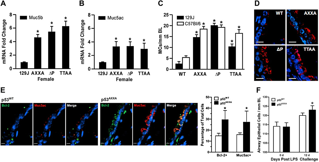
Figure 6. The proline-rich domain of p53 is crucial in regulating mucous differentiation.
RNA from the right cranial lung lobes of p53WT, p53AXXA and p53ΔP mice were analyzed by qRT-PCR for Muc5b (A) and Muc5ac (B). (C) Mucous cells per mm basal lamina were quantified in the axial airways of p53WT, p53AXXA, p53ΔP, and p53TTAA mice on 129J and C57Bl/6 backgrounds. Bars = group means ± standard error from the mean (n = 6 mice/group). * denotes statistically significant difference. (D) Representative micrographs of airway tissue sections from p53WT, p53AXXA, p53ΔP, and p53TTAA mice on 129J immunostained for Muc5ac (red) and counterstained with DAPI (blue) for nuclear staining. Scale - 10 µm. (E) p53WT and p53AXXA mice were instilled with LPS and changes in Bcl-2- and Muc5ac-positive cells was quantified after 12d of instillation. Representative micrographs from p53WT and p53AXXA mice airway sections at 12 d post LPS instillation showing the Bcl-2 (green) and Muc5ac (red) immunostained cells and with DAPI (blue) nuclear counterstaining. Scale - 5 µm. (F) Total airway epithelial cell numbers per millimeter basal lamina at 0 d (baseline) and at 12 d post LPS instillation. Bars = group means ± standard error from the mean (n = > 3 mice/group); * P<0.05.

