Figure 6.

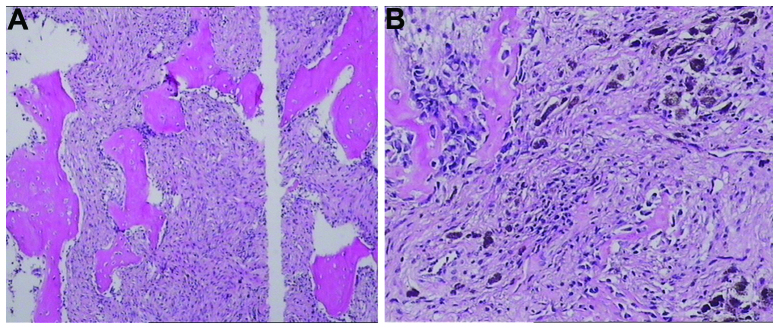
Tumor cell invasion into the surrounding sclerotin. (A) Tumor cells separate from the bone trabecula (magnification, ×400). (B) Tumor cell invasion of the bone trabecula (magnification, ×40) (stain, hematoxylin and eosin).

Tumor cell invasion into the surrounding sclerotin. (A) Tumor cells separate from the bone trabecula (magnification, ×400). (B) Tumor cell invasion of the bone trabecula (magnification, ×40) (stain, hematoxylin and eosin).