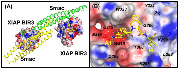
Figure 3.

(A) Crystal structure of the dimeric Smac protein in complex with two XIAP BIR3 proteins (PDBID: 1G73). The AVPI motifs are shown in ball models. (B) Detailed interactions between the AVPI binding motif and XIAP BIR3 residues. Oxygen and nitrogen atoms are colored in red and blue colors. Hydrogen bonds are depicted in dash lines. Electrostatic surfaces of XIAP BIR3 are shown where the red, grey and blue colors denote negative, neutral and positive charged regions. The figures are prepared using the PyMOL and APBS programs.
