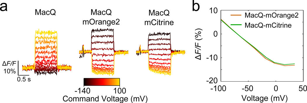
Figure 3. FRET-opsin sensors report voltage depolarization via decreases in emission intensity from the fluorescence donor.
(a) Optical step responses of cultured HEK293T cells transfected with MacQ, and cultured neurons transfected with MacQ-mOrange2 and MacQ-mCitrine (440 Hz frame rate). The MacQ VSD increased its fluorescence intensity with increased voltage depolarization (left). The donor fluorescent proteins of the FRET-opsin sensors exhibited decreases in fluorescence intensity with increased depolarization (middle, right). We held neurons at −70 mV at the start of each trace and stepped to command voltages ranging from −140 mV to +100 mV.
(b) Steady state fluorescence responses of MacQ-mOrange2 and MacQ-mCitrine as a function of neuronal membrane voltage.
Illumination at the specimen plane was 1400 mW mm−2 (λ = 633 nm) for the MacQ studies, and 15 mW mm−2 (λ = 530 nm and 505 nm, for MacQ-mOrange and MacQ-mCitrine, respectively) for the FRET-opsin sensors. Error bars are s.e.m.

