
Figure 1.
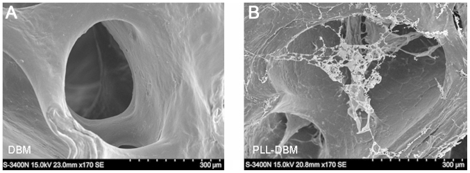
Electron microscope images of (A) DBM and (B) PLL-DBM. The PLL-DBM surface and inner wall of mesh were covered by a milky white PLL coat, and the PLL formed a spider web-like mesh structure in the interspaces. DBM, demineralized bone matrix; PLL-DBM, DBM coated with poly-L-lysine.