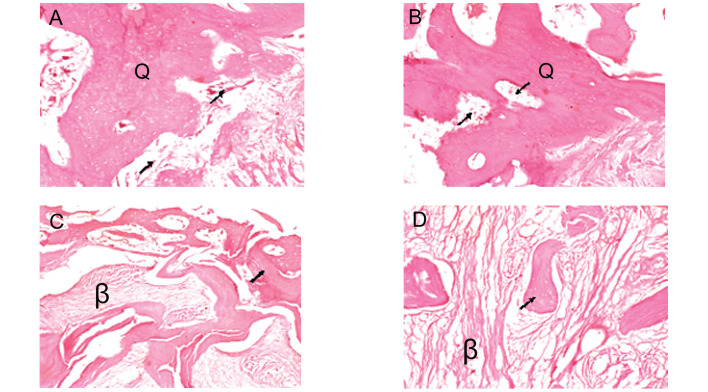
Figure 7.
Histological micrographs (hematoxylin and eosin staining; magnification, ×40). A number of woven bones (Q) had formed and marrow-like tissue (→) was observed in the (A) IA and (B) IB groups; (C) ossification around the graft was active (→), but a number of fibrous tissues (β), which had not yet formed bone, were observed in the center in the IIC group; (D) fibrous tissue (β) was the predominant feature in the IID group, with only a few small, piece-like bones.

