Figure 4.
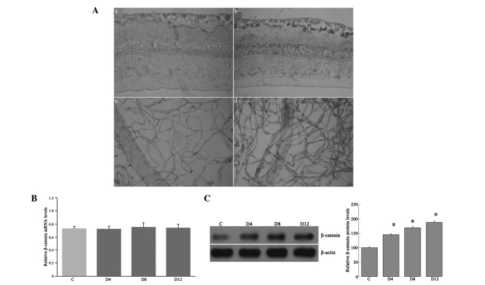
(A) Representative immunohistochemical analysis of β-catenin in the retinas and retinal vasculature. β-catenin immunoreactivity was observed in the retinal vasculature and in the outer plexiform, inner nuclear, inner plexiform and ganglion cell layers of retinas from (a and c) control rats and (b and d) diabetic rats 12 weeks after the induction of diabetes; immunoreactivity was increased in the retinas of the diabetic rats. In the negative control staining, no immunoreactivity for β-catenin was found in the retinas and retinal vasculature (figure not shown) (original magnification, ×400). (B) Reverse transcription-quantitative polymerase chain reaction analysis of β-catenin mRNA expression in the retinas of streptozotocin-induced diabetic and control rats. The retinal β-catenin mRNA expression was examined in the rat retinas at four, eight and 12 weeks after the induction of diabetes (D4, D8 and D12, respectively), as well as in the controls, using β-actin as an internal control. (C) Western blot analysis for β-catenin from the retinal lysates of the rats. Autoradiography depicting the β-catenin shows the β-catenin expression increased in the retinas at four, eight and 12 weeks after the induction of diabetes compared with that in the controls. *P<0.05 vs. the control.

