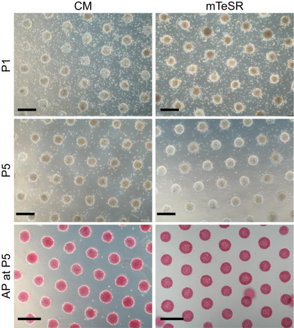
Fig. 1.
Bright-field images of a patterned array of hPSC colonies cultured in conditioned medium (CM, left) and mTeSR (right) after one passage (P1, top) and after five passages (P5, middle). Images were taken at day 2 of culture prior to feeding the cells. The colonies stain positive for alkaline phosphatase (AP) in both CM and mTeSR at P5 (bottom), indicating that the hPSCs have remained pluripotent. The patterned hPSC colonies displayed in this figure are comprised of hESCs from line H9 (WiCell). Scale bars = 400 µm.

