Figure 2(A-C).

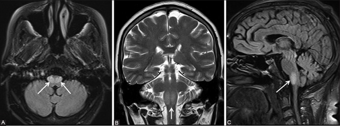
A panel of (A) FLAIR axial (B) T2-weighted coronal, and (C) FLAIR sagittal MRI images showing the hypertrophy of bilateral medullary olives (arrows). Hyperintensity is also noted in the enlarged olives

A panel of (A) FLAIR axial (B) T2-weighted coronal, and (C) FLAIR sagittal MRI images showing the hypertrophy of bilateral medullary olives (arrows). Hyperintensity is also noted in the enlarged olives