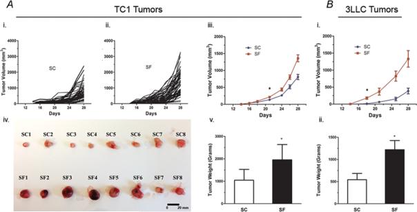
Figure 1. SF induces accelerated tumor size growth both in TC1 and 3LLC tumors.
A. C57/B6 mice exposed to SF starting 1-week prior to flank injection with TC-1 cells showed accelerated tumor size growth and weight with significant differences when compared to mice under SC conditions. (i). shows the trajectory of tumor growth measured under SC conditions (n=53), compared to tumors from SF exposed mice; (ii). SF, n= 55.; (iii). When comparing the average tumor growth for both conditions, statistically significant differences emerged at day 23 following injection and sustained thereafter (p< 0.007). (iv) Macroscopic differences between the SF and SC tumors in samples of enucleated tumors. (v). Tumor weight was also significantly higher at day 28 in SF-exposed mice (SF-57/B6: 1.955± 0.680 g vs. SC-C57/B6: 1.046±0.479 g; p<0.001; n= 109 in each group). Of note, those experiments were performed in batches of 20 mice each, using the same tumor cell injection and follow-up protocols.
B. Mice (n=15/group) exposed to SF and injected with 3LLC cells showed significantly accelerated tumor growth compared to SC conditions, (i) with differences emerging after 19 days from cells injection (p<0.001), and (ii) significant tumor weight differences at enucleation (SF-57/B6: 1.22 ± 0.281 g vs. SC-C57/B6: 0.543 ±0.163 g; p<0.027). *p< 0.05

