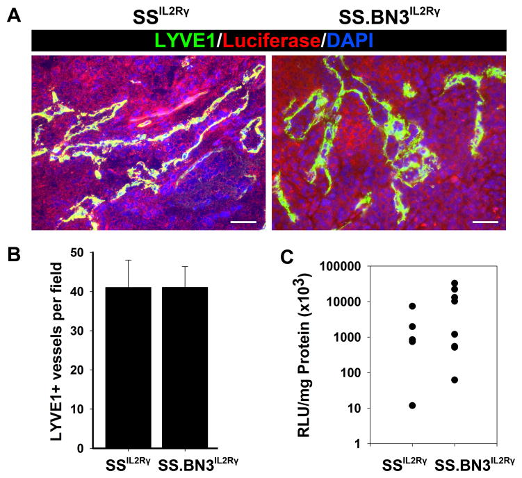
Figure 5.
Characterization of tumor-associated lymphatic vessels and lymphogenous metastasis in 231Luc+ tumors implanted in SS.BN3IL2Rγ and SSIL2Rγ rats at 24 days post-implantation.(A) Visualization of tumor-associated lymphatic vessels at using anti-LYVE-1 staining of 231Luc+ tumors implanted in SS.BN3IL2Rγ and SSIL2Rγ rats. Scale bar represents 100 μm. (B) Mean lymphatic vessel density in 231Luc+ tumors implanted in SS.BN3IL2Rγ and SSIL2Rγ was calculated from three images per tumor (n= 5–6 per strain) acquired at 100X magnification. Data are presented as the mean vascular density per 100X field ± SEM. (C) Lymphogenous metastatic burden was measured by luciferase activity normalized to total milligrams of protein in axillary LN lysates from SS.BN3IL2Rγ (n = 5) and SSIL2Rγ (n = 8) rats. Metastatic burden of individual rats are represented by the dots and the black bars indicate the average metastatic burden per strain. For (B) and (C), statistical analysis of data was performed by Student unpaired t test.

