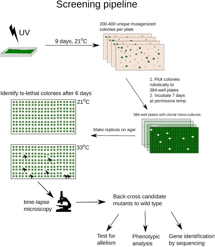
Figure 1.
Screening Pipeline.
UV-mutagenized cells were deposited on agar to form colonies and picked robotically into 384-well plates. After replica pinning, ts mutants were identified on the 33°C plate (black arrowheads) based on reduction of biomass compared with 21°C. All ts mutants were screened by time-lapse microscopy to identify potential cell cycle mutants (div/gex; see text). Candidate div and gex mutants were backcrossed to the wild-type parent and analyzed genetically and phenotypically.
[See online article for color version of this figure.]

