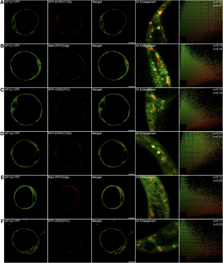
Figure 6.
Subcellular Localization of AP1 Gamma Adaptin.
AP1γ1-YFP ([A] to [C]) and AP1γ2-YFP ([D] to [F]) partially colocalized with the TGN marker RFP-SYP61, while they separated from the Golgi marker ManI-RFP and the PVC marker RFP-VSR2. The right column shows the scatterplot images indicating the extent of colocalization with the linear Pearson correlation coefficient (rp) and the nonlinear Spearman correlation coefficient (rs). Bars = 10 μm.

