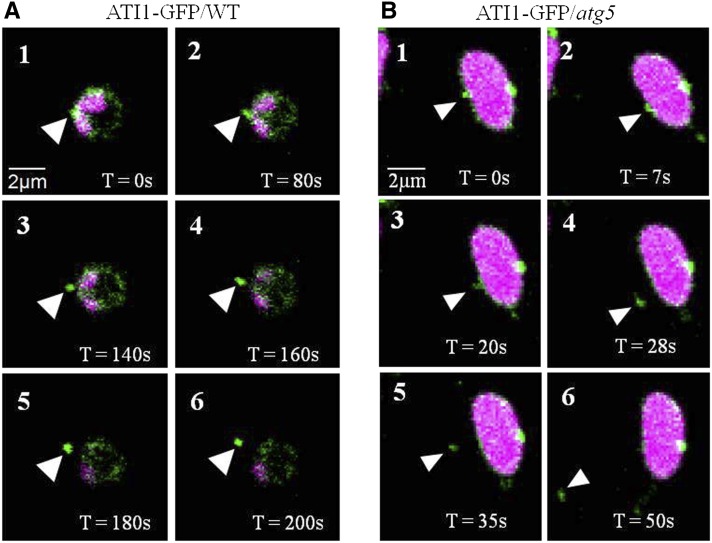
Figure 3.
The Budding and Detachment of ATI-PS Bodies from Plastids.
Confocal time-lapse imaging of plastids from hypocotyl epidermis cells of either an ATI1-GFP transgenic plant (wild-type background; [A]) or a transgenic plant expressing ATI1-GFP in the background of the atg5 autophagy-deficient mutant (B). Both plastids display chlorophyll autofluorescence (magenta signal) and ATI1-GFP expression (green signal). Numbers 1 to 6 (in both panels) represent a series of time points (T) as indicated, and the budding ATI-PS bodies are indicated by white arrowheads.

