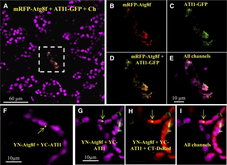
Figure 8.
ATG8f and ATI1 Interaction Is Associated with Plastids in Leaf Mesophyll Cells.
(A) to (E) Confocal imaging of adult rosette leaf mesophyll cells of a transgenic plant stably expressing both mRFP-ATG8f (red signal) and ATI1-GFP (green signal).
(A) A representative image showing a senescing cell (highlighted within the dashed white rectangle) exhibiting a relative low level of chlorophyll emission and expression of both the red and green signals, resulting in yellow color where colocalization occurs. This cell is surrounded by a population of “vital” cells exhibiting no detectable expression of the fluorescent proteins.
(B) to (E) Enlargements of the boxed area in (A).
(F) Confocal imaging of a BiFC assay (see Methods) involving coexpression of YC-ATI1 and YN-ATG8f. The yellow signal indicates interaction between the two proteins.
(G) to (I) A similar BiFC assay as shown in (F) except that the interaction here is presented as green signal and with the additional expression of the CT-DsRed (red) plastid stroma fluorescent marker. In (F) to (I), yellow arrows point toward bodies where the interaction between ATG8f and ATI1 occurs.

