Figure 1.

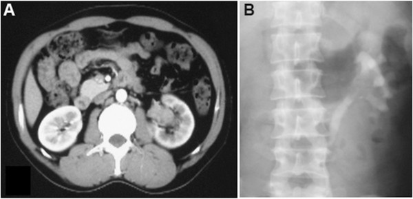
Contrast-enhanced CT demonstrating a 4-cm soft-tissue mass occupying the dilated left renal pelvis. (A) Enhanced CT. (B) Left retrograde pyelography.

Contrast-enhanced CT demonstrating a 4-cm soft-tissue mass occupying the dilated left renal pelvis. (A) Enhanced CT. (B) Left retrograde pyelography.