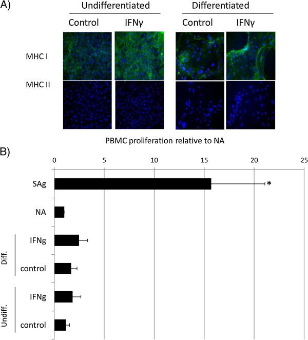
Figure 1.

Proliferation of equine PBMCs is not induced by co-culture with equine ESCs. (A) Immunocytochemical staining of IFN-γ-treated embryo-derived stem cells (ESCs) for MHC I and MHC II. Cell nuclei are indicated by blue Dapi staining, and expressed MHC I or II proteins, by green staining. Representative images from one of three replicates are shown. (B) Relative proliferation of peripheral blood mononuclear cells (PBMCs) to undifferentiated (ESCs) and differentiated (dES) ESCs cultured in the presence and absence of IFN-γ, where NA is baseline, nonactivated PBMC proliferation; sAg is superantigen-stimulated PBMCs (positive control); IFN-γ is 72-hour pretreated undifferentiated or differentiated ESC. *Results significantly different relative proliferation when compared with NA PBMCs (P < 0.05). Error bars represent the standard error of seven individual experimental repeats using three different cell lines.
