
Figure 2.
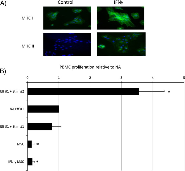
Proliferation of equine PBMCs is suppressed by co-culture with equine MSCs. (A) Immunocytochemical staining of IFN-γ-treated mesenchymal stem cells (MSCs) for MHC I and MHC II. Cell nuclei are indicated by blue Dapi staining, and expressed MHC I or II proteins, by green staining. Data show representative images from one of three replicates. (B) Relative proliferation of effector (Eff#1) peripheral blood mononuclear cells (PBMCs) to mesenchymal stem cells (MSCs) cultured in the presence and absence of IFN-γ. Autologous and allogeneic stimulator cells are used as negative and positive controls (Stim #1 and Stim #2, respectively). *Results significantly different from those for nonactivated (NA) PBMCs (P < 0.05). Error bars represent the standard error of six individual experimental repeats by using three different cell lines.