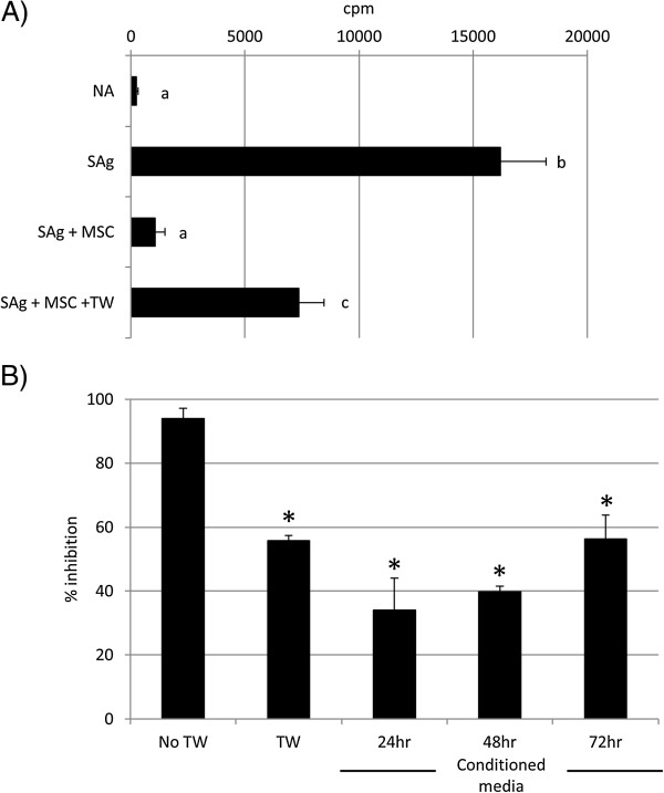
Figure 5.

Soluble factors are involved in MSC-mediated PMBC suppression. (A) Mesenchymal stem cells (MSCs) suppress S. equi superantigen (sAg)-induced peripheral blood mononuclear cells (PBMCs) proliferation when separated by a transwell membrane (TW). Graph depicts radioactive thymidine (3H-thymidine) counts per minute (cpm) as a measure of cell proliferation. Differing letter annotations denote a significantly different mean (ANOVA, all P < 0.05). Error bars represent the standard error of the mean of three biologic repeats. (B) Exposure to 24-, 48-, and 72-hour mesenchymal stem cell (MSC)-conditioned media suppresses S. equi superantigen (sAg)-induced peripheral blood mononuclear cell (PBMC) proliferation, but to a lesser extent than via direct cell-to-cell contact. TW, transwell. *Results significantly different from no transwell (No TW) values (ANOVA; P < 0.05). Error bars represent the standard error of the mean of three biologic repeats.
