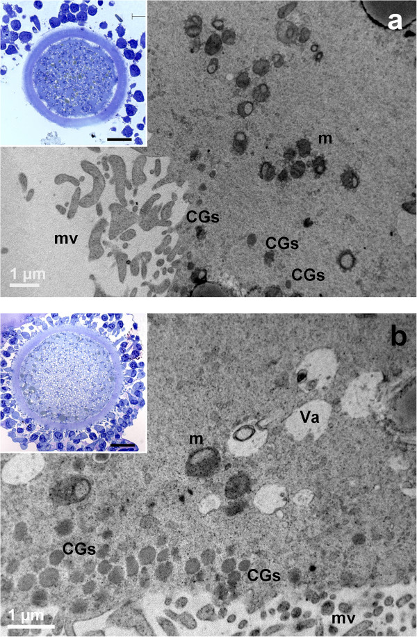
Figure 6.

General morphology and organelle microtopography in prepubertal (a) and adult (b) ovine cumulus-oocyte-complexes (COCs) after 19 hours of IVM. a) Representative TEM micrograph showing an oocyte cortex with mitochondria (m), provided by numerous cristae. Cortical granules (CGs) are irregularly distributed in the sub-plasmalemmal region; mv: microvilli. Bar: 1 μm. Inset in a): representative LM image showing spikes of the oolemma, associated with CC detachment. Bar: 20 μm. b) Representative TEM micrograph showing an ooplasm with several irregularly shaped vacuoles (Va), mitochondria (m) and multiple layers of electron-dense cortical granules (CGs). Bar: 1 μm. Inset in b): representative LM image showing a regularly round shaped oolemma; mv: microvilli. Bar: 20 μm.
