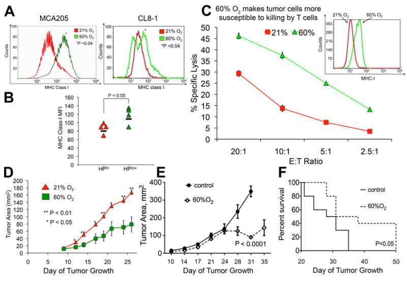
Fig. 4. Hyperoxic breathing increases tumor cell recognition and killing by antitumor T cells, enhances tumor regression, and improves survival of tumor-bearing mice.

(A) Expression of antigen-presenting MHC class I molecules on the surface of MCA205 fibrosarcoma and B16 melanoma tumors (H2Kb transfected B16, CL8-1 line) Mean fluorescent intensity of MHC-I expression was measured by flow cytometry in mice breathing 60% or 21% oxygen (MCA205, P < 0.04, n = 3; CL8-1, P < 0.04, n = 4). (B) Tumor cells exposed to higher levels of hypoxia had lower surface expression of MHC class I (P < 0.05, n = 4). (C) Hyperoxia is increases lethal hit delivery by cytotoxic T lymphocytes. MCA205 cells cultured in 21% or 60% oxygen for 4 days in vitro demonstrated an increase in the expression of MHC-I as measured by flow cytometry (inset, P < 0.01). The cells were 51Cr-labeled and used in a cytotoxicity assay with culture-activated T cells from MCA205 tumor draining lymph nodes at different effector/target (E/T) ratios. After 4 h, radioactivity released from lysed target cells was counted on a γ-counter (*P < 0.01). (D) Tumor regression of B16 (H2Kb transfected B16, CL8-1 line) tumor-bearing mice following 60% oxygen breathing. After s.c. tumor establishment identified by the first appearance of palpable tumors, mice were placed in either 21% or 60% oxygen and tumor size was measured in two dimensions (*P <0.0.5, **P< 0.01, n = 5 mice). (E) Tumor regression of B16 melamona tumors following 60% oxygen breathing. Mice bearing established B16 s.c. melanomas were placed in either 21% or 60% oxygen and tumor size was measured in two dimensions (P < 0.0001, n = 10). (F) Prolonged survival in mice with established B16 tumors breathing 60% oxygen. Mice bearing established B16 s.c. melanomas were placed in either 21% or 60% oxygen. Tumor-bearing mice were euthanized when tumor reached 2 cm in one direction (P < 0.05, n = 10).
